Tadalafil zeigt eine konstante Resorption im Gastrointestinaltrakt, mit maximalen Plasmaspiegeln nach rund zwei Stunden. Der Wirkstoff verteilt sich gut im Gewebe und weist eine hohe Plasmaproteinbindung auf. Seine lange Halbwertszeit erlaubt eine verlängerte Wirkphase. Der Metabolismus erfolgt über das hepatische Enzymsystem CYP3A4, mit der Bildung inaktiver Metaboliten. Exkretion geschieht primär über den Stuhl. Die Häufigkeit von Nebenwirkungen steigt mit der Dosis, wobei vor allem vasodilatatorische Effekte dominieren. Ein gängiger Bezugspunkt in pharmakologischen Analysen ist cialis ohne rezept, das mit dieser Wirkstoffklasse assoziiert ist.
Eujapan.co.jp
Healing of Neuropathic Foot Ulcer using a Novel ‘Wound Boot’ (Kerraboot™)
Leigh R1, Latif N1, Hollingsworth S2, Barker S2, Hurel SJ1
INITIAL MANAGEMENT OF PATIENT'S HEEL ULCERS
Department of Diabetes1 and Surgery2, University College Hospitals, London,
IV antibiotics (ceftazidime, flucloxacillin, ciprofloxacin, metronidazole)
Wound care (dry dressings changed every 2 days)
Abstract: A 67-year-old man, with poorly controlled Type 2 diabetes, developed
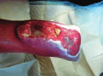
ulceration of both heels during a prolonged hospital admission for septicaemia. The left heel ulcer measured 8cm in maximum diameter and was approximately 1 cm deep (Figure 1). The right heel ulcer measured 3cm in maximum diameter
and was approximately 0.3cm in depth. He had a peripheral sensory neuropathy.
Doppler arterial ultrasound gave abnormally high ankle-brachial pressure indices,
secondary to vessel calcification. Plain radiographs of the foot were suggestive of
underlying osteomyelitis of the calcaneum. Magnetic resonance and nuclearimaging supported this diagnosis.
Despite bed rest, wound toileting and appropriate antimicrobials, the left heel
ulcer continued to deteriorate with ulceration and infection spreading proximally
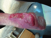
to involve the posterior tibial compartment. To circumvent amputation,
considered at the time to be the only suitable procedure, a novel ‘wound boot’ was employed. The boot has been designed to provide a highly cost-effective, carer-friendly alternative to traditional dressings, whilst providing an optimal
environment for wound healing. The boot was changed twice daily, often by the
patient himself, and used in total for three weeks. Over this period, the infectionresolved completely and the wound showed dramatic signs of healing (Figure 2). INTRODUCTION
Diabetic neuropathic ulceration is extremely difficult to treat. Ischaemia andinfection, affecting superficial tissues or the underlying bone, frequentlycomplicate the condition. The management of these chronic ulcers ismultidisciplinary; requiring scrupulous wound hygiene, careful choice and regular
MANAGEMENT OF LEFT HEEL ULCER WITH KERRABOOT
changing of dressings, systemic antibiotic therapy, and limb rest. Despite intensivemanagement, healing time is frequently protracted and success rates are poor. The
The Kerraboot was applied twice daily for 24 days.
management of chronic diabetic foot ulcers makes considerable demands on
The patient was easily able to change the boot, after simple instruction.
nursing time, in hospitals and the community. [It has been estimated that over
25% of the district nurse’s workload involves ulcer care.]
KERRABOOT
The Kerraboot ‘wound boot’ (Ark Therapeutics) is a tri-laminar, foot-shaped
plastic boot, which fastens securely around the leg to enclose the area of
ulceration. The key elements of the Kerraboot are: • Warm, moist, protected environment – to promote wound granulation and
ulcer margins contract and granulation at base of ulcer; CRP 12.7
• Super-absorbent padding – to remove excess moisture and wound exudate,
• Integral; charcoal filter and carbon impregnated material – to eliminate odour.
• Textured base – to prevent slipping during patient mobilisation. CASE REPORT • Patient – male, 67 years.
– with weekly review at diabetes podiatry clinic. MEDICAL HISTORY
Antibiotics – rifampicin and fusidic acid.
• 1983 Type 2 diabetes – glibenclamide (10 mg o.d.)
• Poor glycaemic control – glycosylated haemoglobin 12.5% (normal 4-6%)
• Regular alcohol consumption – 6 units daily
POTENTIAL BENEFITS OF THE KERRABOOT FOR DIABETIC FOOT ULCERS
• 1987 progressive neuropathy – initially left foot, then both feet
• Other diabetic complications – proliferative retinopathy and nephropathy
• Warm, moist, protected environment to promote wound healing
• 1996 CVA – persistent left hemi-paresis
• Super-absorbent material to absorb exudate and excessive moisture
• June 1999 – Gram-negative septicaemia (secondary to UTI) - prolonged
• Transparent material to allow easy monitoring of the ulcer
• Efficient odour control to improve patient and ward acceptability
• Development of pressure sores to heels – discharged, ulcer care by
• Ease of use – patient or carer can change Kerraboot with minimum of
• October 1999 significant deterioration of both ulcers – re-admitted to hospital
• Potential cost benefits from reduced hospitalisation and nursing workload
• Opportunity to increase patient involvement in wound management. INVESTIGATIONS Both ulcers produced a brown, foul-smelling exudate. The right ulcer was 3cm in diameter and 0.3cm deep; it showed no sign of cellulitis. The left ulcer measured CONCLUSION
approximately 8cm in diameter and was 1cm deep. The surrounding tissue wasvery macerated and there was local cellulitis.
This intervention appeared to promote healing of a chronic, infected,necrotic, neuropathic heel ulcer in a diabetic patient, and circumvented the
Doppler arterial ultrasound – high ankle-brachial pressure indices (e.g. Dorsalis
pedis: L >300mmHg, R 240mmHg), probably secondary to vessel calcification.
The evidence from this case suggests that the Kerraboot produces an
Microbiology – initial wound cultures grew Pseudomonas aeruginosa and
environment conducive to wound healing. A study is currently underway to
Radiography, MR and nuclear imaging – osteomyelitis of calcaneum.
Biochemistry – C-reactive protein (CRP) 94, erythrocyte sedimentation rate (ESR)80, white cell count (WCC) 8.3 x 10⁹/L.
S u p p o r t e d by a n e d u c a t i o n a l g ra n t f ro m A r t Th e ra p e u t i c s
6 Wa r re n M ew s , L o n d o n W 1 T 6 A R Te l e p h o n e . 0 2 0 7 3 8 8 7 7 2 2
Public Health Advancement of global health: key messages from the Disease Control Priorities Project Ramanan Laxminarayan, Anne J Mills, Joel G Breman, Anthony R Measham, George Alleyne, Mariam Claeson, Prabhat Jha, Philip Musgrove, Jeff rey Chow, Sonbol Shahid-Salles, Dean T Jamison The Disease Control Priorities Project (DCPP), a joint project of the Fogarty International Center o
RSV – Recommended Literature Down Syndrome/Trisomy 21 • Aboussouan LS, O’Donova PB, Moodie DS, Gragg LA, Stoller JK Hypoplastic trachea in Down’s Syndrome. Am Rev Resp Dis, 1993; 147: 72-5. • Bloemers BL, Bont L, de Weger RA, Otto SA, Borghans JA, and Tesselaar K. Decreased Thymic Output Accounts for Decreased Naïve T Cell numbers in Children with Down Syndrome. J Im

 Healing of Neuropathic Foot Ulcer using a Novel ‘Wound Boot’ (Kerraboot™)